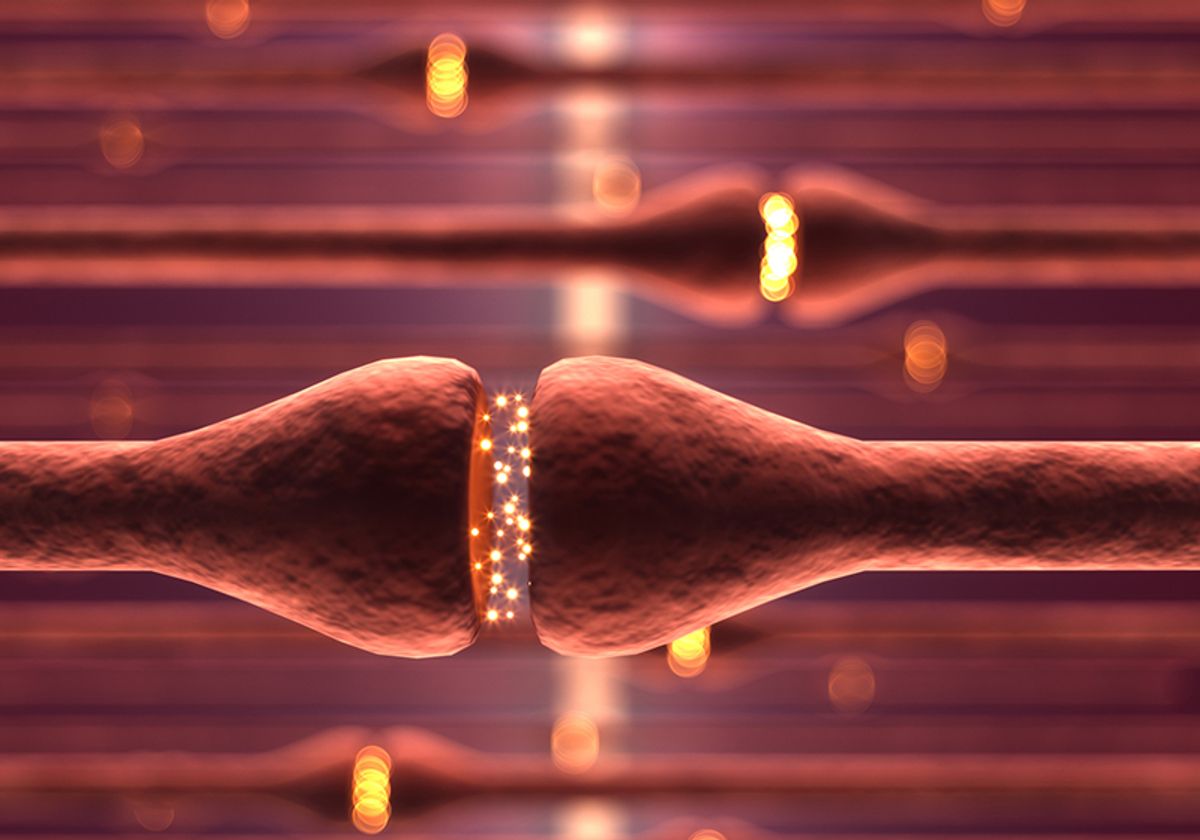
The human body is a dynamic vessel of sensors and transducers that translate messages from external stimuli into the electrical and chemical language of the brain. Among the human sense organs, the eye is an elegant model of phototransduction, converting light into the electrical energy that powers vision. Specialized cells in the retina, known as photoreceptor cells, detect light and relay this information to the brain through a multilayered chain of neurons. These neurons communicate across synapses—the physical gaps between nerve cells that serve as transmission hubs.
Researchers use human pluripotent stem cells to create retinal organoids—3D cellular models of the retina—to study eye development, normal vision, and degenerative diseases. They also use these organoids to produce retinal neurons that may one day replace damaged or diseased cells in the eye. The cells in these organoids form synapses and communicate with one another, but scientists must disassemble organoids into individual cells to produce retinal neurons. This necessary disruption breaks down synaptic connections and affects how the neurons function and communicate.
Researchers from the University of Wisconsin-Madison recently examined whether retinal organoid neurons derived from human pluripotent stem cells can still form synapses with one another and maintain connectivity after organoid disassembly—a necessary prerequisite for retinal neural circuit function.1 They used inactivated rabies virus, which is known to pass from one neuron to another by synaptic transmission,2 to label and count retinal neurons.
The researchers found that the dissociated retinal organoid neurons retained the ability to form synaptic connections, based on the movement of the labeled virus from one cell to the next. “What was surprising was the efficiency with which that would happen,” said David Gamm, a pediatric ophthalmologist, research professor, and lead author of this study. “Photoreceptors were the most prominent population of cells that could do that. I thought these may be rare events, of which photoreceptors would be a minority population, if present at all. Instead, it was one of our majority populations.”

“The approach is robust and reproducible,” said Maksim Plikus, a professor of developmental and cell biology at the University of California, Irvine, who was not involved in this study. He explained that treating retinal degeneration using transplanted retinal neurons has the potential to restore vision or stop disease progression. “While derivation of new retinal neurons, including photoreceptors, is now quite efficient and reproducible, it remains an open question to which degree such grafted neurons integrate into existing retinal networks and [whether] they form functional synapses,” said Plikus.
Gamm is excited to answer these questions as he sets his sights on future clinical studies in patients. Data from in vitro experiments and animal models are indispensable but limited. “There's only so much you can do with graphs. At some point, you've got to do that human experiment. We realized collectively that we're at a point where we just won't know until we start doing rigorous studies, seeing what happens, learning, and improving,” said Gamm.
The results of Gamm’s latest study are the culmination of a decade-long retinal organoid research program, the foundation of which rests on basic developmental biology. “Retinal organoids provide us insight into how the human retina develops, which in turn tells us how we might be able to repair it over time,” Gamm said. This potential repair process carefully considers how the packets of energy carried by light particles are absorbed by the retina and converted into electrical signals that enable vision. It is by reflecting on how humans experience the codes hidden in the world’s sensory messages that retinal stem cell therapy will advance from a vision to reality.
References
- Ludwig AL, et al. Re-formation of synaptic connectivity in dissociated human stem cell-derived retinal organoid cultures. PNAS. 2023;120(2):e2213418120.
- Beier KT. The serendipity of viral trans-neuronal specificity: more than meets the eye. Front Cell Neurosci. 2021;15:720807.


